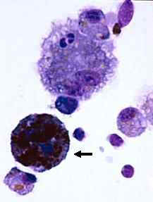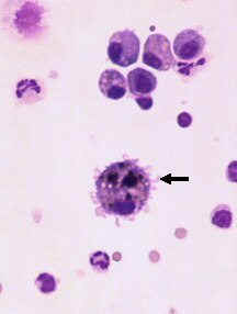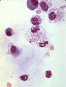
Wright's stain, 500x
The macrophage close to the center of this image has phagocytized a red blood cell which is located in a cytoplasmic vacuole. It is important to differentiate erythrophagocytosis from a red blood cell that happens to be resting above a macrophage. In fluid samples, erythrophagocytosis can also occur in vitro. The presence of erythrophagocytosis suggests recent hemorrhage. With time, the degradation of hemoglobin will be associated with the formation of a brown-black pigment, called hemosiderin, which will eventually become yellow or orange (hematoidin). These pigments can be found inside macrophages or extracellularly.