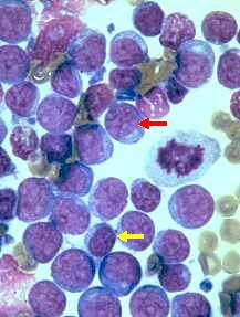
Lymphosarcoma: Wright's stain, 1000x
Malignant
lymphomas, or lymphosarcomas are frequently (but not always!) characterized by the
predominance of immature lymphocytes, which eventually replace the normal lymphoid
population. A diagnosis of malignant lymphoma can be made based on cytology when immature
lymphocytes account for more than 50% of the cell population. However, it is possible for
neoplastic cells to account for less than 50% of the cellular population, making it
difficult to make a diagnosis based on cytology. Generally, neoplastic immature lymphoid
cells may be larger than neutrophils, possess granular chromatin, have visible nucleoli
(red arrow), and have a nucleus to cytoplasm ratio that is lower than that of a mature
lymphocyte (yellow arrow). Also note the presence of mitotic figures in this
microphotograph. It is important to remember that the observation of mitotic figures alone
does not indicate a malignant change. In the case of a well differentiated lymphoma (or
lymphoma comprising small lymphocytes), histology will generally be necessary for a
diagnosis. This can also be said of any cytology of an enlarged lymph node presenting a
predominance of mature lymphocytes without an obvious inflammatory component or increased
number of plasma cells to suggest lymphadenitis or lymph node hyperplasia, respectively.
In such a case, the architecture offered by histopathology will allow for a more definite
diagnosis.