Erythrocyte agglutination
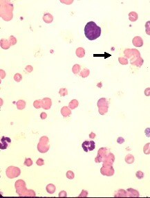 Wright's
Stain, 500x |
Erythrocyte
agglutination observed on a blood smear
The observation of red blood cell agglutination
(also referred to as autoagglutination) must be distinguished from rouleaux formation which is a physiological phenomenon. The
presence of antibodies (usually IgM) on the surface of red blood cells is responsible for
the phenomenon of autoagglutination. Agglutination can be observed during immune-mediated
hemolytic anemia, but also during 'cryoglobulinemia' ( a far more rare condition).
Agglutinating red blood cells resemble grapelike clusters whereas red
blood cells in rouleaux formation resemble a stack of coins.
In order to clearly distinguish erythrocyte agglutination from
rouleaux formation, a simple saline test can be performed. |
Return
to menu
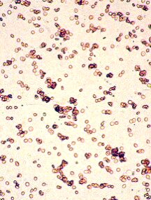
Saline Test
This test confirms the presence of agglutination by mixing a drop of
uncoagulated blood with a (larger) drop of saline solution. The mixture is then placed on
a slide with a coverslip and observed under the microscope. In the presence of
agglutination, the red blood cells will remain clumped: this indicates a positive test . In the presence of rouleaux formation (a
physiological phenomenon associated with plasma proteins), the red blood cells will spread
out individually: this indicates a negative test .
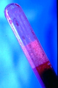
Agglutination, if severe enough, may be observed macroscopically by
noting the blood running along the inner wall of the purple-top tube.
Positive saline test, 100x
Negative saline test, 100x
Return to
menu