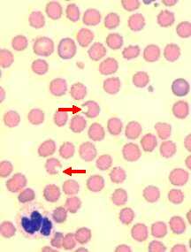
Wright's stain, 1000x
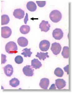
Acanthocytes (red arrow) and schizocytes (black arrow) in a canine blood smear
The observation of acanthocytes (red arrow), schizocytes (black arrow), or keratocytes on a blood smear suggests the presence of microangiopathy which has resulted in intravascular fragmentation of red blood cells. If severe enough, erythrocyte fragmentation may contribute to the development of anemia (this would be one of the types of hemolytic anemia). Erythrocyte fragmentation can be seen in several conditions affecting blood vessels including disseminated intravascular coagulation, vasculitis or other processes that involve a vascular anomaly, such as hemangiosarcoma. This neoplasm of blood vessels (often but not exclusively involving the spleen and/or liver) is not uncommon in older, large breed dogs. Naturally, erythrocyte fragmentation is not diagnostic for this tumor. However, the observation of erythrocyte fragmentation, especially in an older dog of a large breed (ex: German Shepherd), should lead one to consider hemangiosarcoma as part of the differential diagnosis. Finally, although the observation of keratocytes and schizocytes on a blood smear more specifically call to mind the possibility of erythrocyte fragmentation in the patient, the observation of acanthocytes is not specific to this process. Acanthocytes may be formed subsequent to alterations in the composition of the cell membrane in association with liver disease, for instance. Furthermore, a small number of acanthocytes on a blood smear may not be clinically significant.