Faculté de médecine vétérinaire
Département de pathologie et microbiologie

 |
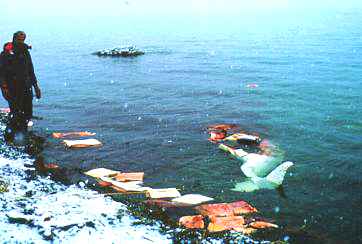
Saint Lawrence belugas Lesions and contaminants
|
||||||||||||||||||||||||
One adult animal out of 5 suffered from cancer. (Cancer was observed in 27 % of examined adult animals from 1983 to 1998). In the western world, cancer causes 23 % of all deaths in humans, a percentage similar to that found in Saint Lawrence beluga. Such a high percentage had never been observed in any wild animal species, terrestrial or aquatic (with the important exception of fish). To our knowledge, this is the first population of wild mammals that can be compared to humans in this regard. The rate of cancer in Saint Lawrence beluga is also much higher than that observed in other cetaceans. Only 28 other cases of cancer have been reported in wild and captive cetaceans worldwide. Thus, cancers reported in Saint Lawrence beluga represent about half of all cancers reported in cetaceans worldwide.
|
|||||
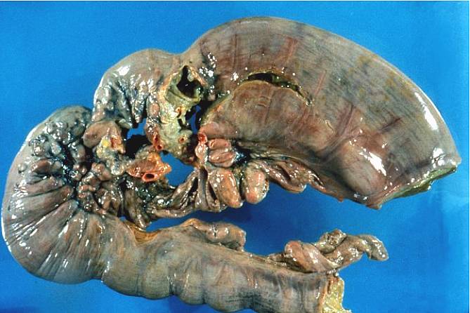 |
Intestinal cancer in a beluga from the St Lawrence Estuary. Examined on May 23, 1993. The larger intestinal segment on the right is abnormally dilated because a malignant tumor (the irregularly sized masses in the center) distorted the intestinal architecture and obstructed the intestinal lumen. Food cannot pass and as a result, accumulates in the intestinal segment that is closest to the stomach (on the right). |
||||
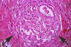 |
Microscopic view of intestinal cancer in an adult beluga whale. The paler area delineated by the two arrows is a nest of tumor cells. Within this paler area, gland-like structures (white spaces) are lined by cancer cells. Cancer cells mimic the glands normally found in the intestinal mucosa. The "glands" are irregular and ill formed, in contrast with normal intestinal crypts. These glandular structures are not present where they normally should be, near the intestinal lumen. Rather, they are embedded deep in the muscular layers, where they destroy vital structures such as blood vessels. | ||||
BibliographyNature Reviews Cancer 9, 605 (August 2009) | doi:10.1038/nrc2698 Science and society : Wildlife cancer: a conservation perspective Denise McAloose & Alisa L. Newton
|
|||||